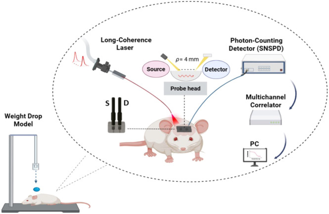
Traumatic brain injury (TBI) can lead to long-lasting impairments in cerebral perfusion, making early detection of microvascular changes critical for guiding clinical interventions. We employed time-domain diffuse correlation spectroscopy (TD-DCS) at 1064 nm to noninvasively quantify depth-resolved cerebral blood flow (CBF) and low-frequency oscillations (LFOs) in a mouse model of closed-head injury. By analyzing earlier photon arrivals (with greater superficial weighting) and later photon arrivals (with enhanced sensitivity to deeper tissue), we identified a significant drop in CBF shortly after injury, with partial recovery observed at 2 h post-trauma. Power spectral analysis of the blood flow index (BFI, a diffusion coefficient proportional to CBF) revealed significant alterations in LFO bands, particularly in slow-5 (0.01 to 0.027 Hz) and slow-3 (0.073 to 0.198 Hz) ranges. These differences, assessed using a paired Wilcoxon rank-sum test (𝑝<0.05), were more pronounced than BFI alterations alone, indicating that LFOs may serve as sensitive biomarkers of neurovascular disruption. Our findings demonstrate the feasibility of TD-DCS for relative depth-sensitive monitoring of cerebral hemodynamics and oscillatory dynamics after TBI and highlight its potential utility in translational neurotrauma research.